Elbow Anatomy:
Joint Type: elbow - hinge joint; proximal radioulner joint - pivot joint. (Seely, VanPutte, & Russo, 2011, p. 254).
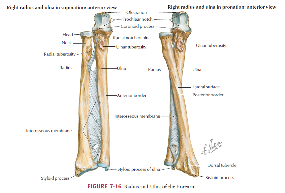
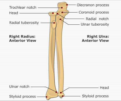
Radial head: The head is on the lateral part of the arm (Hansen, Netter, & Consult, 2010, p.312). The head articulates with the capitellum of the humerus and the radial notch of the ulna (Hansen et al., 2010, p. 312).
Neck & tuberosity (of radius): This area of the radius is a projection along the proximal medial anterior part of the radius (GetBodySmart, n.d.). It is an attachment point for the biceps muscle (GetBodySmart, n.d.). The neck joins the head to the shaft (Anatomy TV, n.d.).
Hansen et al., 2010, p. 427
Styloid process of radius & ulna: The styloid process of the radius is a pointed lateral piece at the distal end of the radius, which forms the lateral portion of the wrist joint (GetBodySmart, n.d.). The radius styloid process rotates around the ulna during forearm rotation (Anatomy TV, n.d.). The styloid process of the ulna is a small, medial projection from the head of the ulna and it forms the medial portion of the wrist joint (GetBodySmart, n.d.).
Ulnar head: The ulnar head is on the medial, distal part of the forearm (Seeley et al., 2011, p. 233). The head articulates with the radius and bones of the wrist (Seeley et al., 2011, p. 233).
Ulnar tuberosity: The proximal end of the ulna is C-shaped and has a trochlear notch and the ulnar tuberosity (Seeley et al., 2011, p .233). This notch fits over the trochlea at the distal end of the humerus and it is held together by the olecranon process and the coronoid process (Seeley et al., 2011, p. 233). The tuberosity is an attachment point for the brachialis muscle (GetBodySmart, n.d.).
Ulnar tuberosity: The proximal end of the ulna is C-shaped and has a trochlear notch and the ulnar tuberosity (Seeley et al., 2011, p .233). This notch fits over the trochlea at the distal end of the humerus and it is held together by the olecranon process and the coronoid process (Seeley et al., 2011, p. 233). The tuberosity is an attachment point for the brachialis muscle (GetBodySmart, n.d.).
Hansen et al., 2010, p. 426
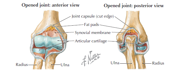
Synovial membrane: this membrane produces synovial fluid which lubricates the joint (Thomas, L. (September 9, 2013). Lecture 1: Orientation to Musculoskeletal Ultrasound [PowerPoint]).
Humerus trochlea: This articulates with the ulna (Seeley et al., 2011, p. 233). It is the medial of the two distal condyles of the humerus (Anatomy TV, n.d.).
Capitulum: The lateral of the two distal condyles of the humerus, it articulates with the radius (Anatomy TV, n.d.).
Humerus trochlea: This articulates with the ulna (Seeley et al., 2011, p. 233). It is the medial of the two distal condyles of the humerus (Anatomy TV, n.d.).
Capitulum: The lateral of the two distal condyles of the humerus, it articulates with the radius (Anatomy TV, n.d.).
Medial and lateral epicondyle: Located on the distal end of the supracondylar ridges of the humerus, they are palpable (Anatomy TV, n.d.).
Medial and lateral supracondylar ridge: Vertical ridges along the medial and lateral aspects of the shaft of the humerus (Anatomy TV, n.d.).
Medial and lateral supracondylar ridge: Vertical ridges along the medial and lateral aspects of the shaft of the humerus (Anatomy TV, n.d.).
GetBodySmart.com
Coronoid fossa: The lateral fossa on the anterior part of the distal end of the humerus that holds the coracoid process of the ulna (Anatomy TV., n.d.).
Radial fossa: The medial fossa found anteriorly on the distal end of the humerus where it holds the head of the radius (Anatomy TV., n.d.).
Olecranon fossa: A deep fossa found posteriorly above the distal condyles of the humerus; it holds the olecranon of the ulna (Anatomy TV., n.d.).
Radial fossa: The medial fossa found anteriorly on the distal end of the humerus where it holds the head of the radius (Anatomy TV., n.d.).
Olecranon fossa: A deep fossa found posteriorly above the distal condyles of the humerus; it holds the olecranon of the ulna (Anatomy TV., n.d.).
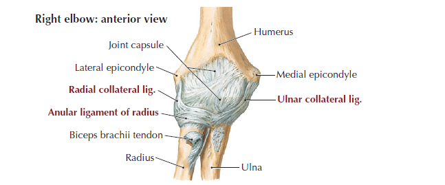
Radial collateral ligament: This ligament is part of the humeroradial joint (Hansen et al., 2010, p. 315). It surrounds the joint, and goes from the lateral epicondyle of the humerus to the radial notch of ulna and anular ligament (Hansen et al., 2010, p. 315).
Annular ligament: This ligament surrounds the radial head and radial notch of the ulna (Hansen et al., 2010, p. 315). It holds the radial head in the radial notch and allows for the movement of pronation and supination (Hansen et al., 2010, p. 315). This is at the proximal radioulner joint (Hansen et al., 2010, p. 315).
Annular ligament: This ligament surrounds the radial head and radial notch of the ulna (Hansen et al., 2010, p. 315). It holds the radial head in the radial notch and allows for the movement of pronation and supination (Hansen et al., 2010, p. 315). This is at the proximal radioulner joint (Hansen et al., 2010, p. 315).
Hansen et al., 2010, p. 426
Lateral ulnar collateral ligament: This ligament extends from the lateral epicondyle to the crista supinator of the proximal ulna where it inserts (Jacobson, 2013, p. 72).
Tendon of the biceps brachii: This tendon inserts on the radial tuberosity (Jacobson, 2013, p. 72).
Brachialis: It originates from the humerus shaft and inserts onto the coronoid process and tuberosity of the ulna (Anatomy TV, n.d.).
Tendon of the biceps brachii: This tendon inserts on the radial tuberosity (Jacobson, 2013, p. 72).
Brachialis: It originates from the humerus shaft and inserts onto the coronoid process and tuberosity of the ulna (Anatomy TV, n.d.).
Olecranon bursa: it overlies the olecranon process on the posterior aspect of the elbow (Anatomy TV, n.d.).
Common extensor lateral: Located on the lateral epicondyle of the humerus and is an attachment site for the extensor carpi radialis brevis, the extensor digitorum, the extensor digiti minimi, and the extensor carpi ulnaris (Anatomy TV, n.d.).
Common flexor origin medial: It originates at the medial epicondyle of the humerus and allows for flexion of the phalanges, metacarpophalangeal joints and wrist (Anatomy TV, n.d.).
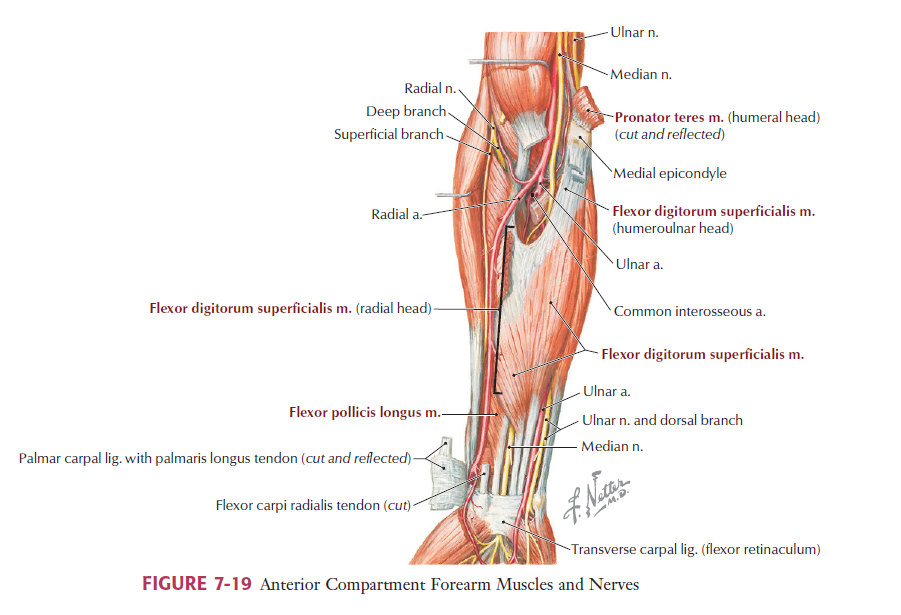
NERVES:
Ulnar nerve:
This nerve runs in the space between the olecranon process of the ulna and the medial epicondyle (Jacobson, 2013, p. 73). Just below this space the ulnar nerve runs through the true cubital tunnel, between the dual origins of the flexor cari ulnaris and deep to the arcuate ligament (Jacobson, 2013, p. 73).
Median nerve:
This nerve is found between the ulner and humeral heads of the pronator teres (Jacobson, 2013, p. 73). It is medial to the brachial artery (Jacobson, 2013, p. 73). It is at the posterior part of the humeral shaft and goes distal and lateral beneath the brachioradialis (Jacobson, 2013, p. 73).
Interosseuous nerve:
It beings anterior to the lateral epicondyle of the humerus and enters the cubital fossa where it goes into the supinator to reach the extensor compartment of the forearm (Anatomy TV, n.d.).
Hansen et al., 2010, p. 436
References:Anatomy.TV StatRef - Interactive Anatomy. (n.d.). Anatomy.tv - Interactive Anatomy. Retrieved October 14 2013, http://www.anatomy.tv/MSKUpperLimb/release/default.aspx?app=mskultrasound_upperlimb
Hansen, J. T., Netter, F. H. 1., & MD Consult LLC. (2010). Netter's clinical anatomy (2nd ed.). Philadelphia: Saunders/Elsevier.
Jacobson, J. (2013). Ch 4 Elbow Ultrasound. Fundementals of musculoskeletal ultrasound (2nd ed., pp. 72 - 85). Philadelphia, PA, USA: Elsevier Inc.
GetBodySmart (n.d.). GetBodySmart: Radius & Ulna Bones | Anterior Markings | Anterior Landmarks | Anterior Anatomy. Retrieved October 14, 2013, from http://www.getbodysmart.com/ap/skeletalsystem/skeleton/appendicular/upperlimbs/radiusulna1/tutorial.html
Seeley, R. R., VanPutte, C. L., Regan, J., & Russo, A. (2011). Ch 10 Muscular System: Gross Anatomy. Seeley's anatomy & physiology (9th ed., pp. 233, 254). New York, NY, USA: McGraw-Hill.
